To elucidate the N-terminal amino acids of a protein, the classic step-wise chemical cleavage by automated phenylisothiocyanate chemistry as developed by Pehr Edman is a straightforward method. If you need to know the identity of a protein, have a look at protein identification by mass spectrometry which is more sensitive and applicable to a wider range of samples. When sample amounts are very limited, mass spectrometry is also worth considering.
We provide
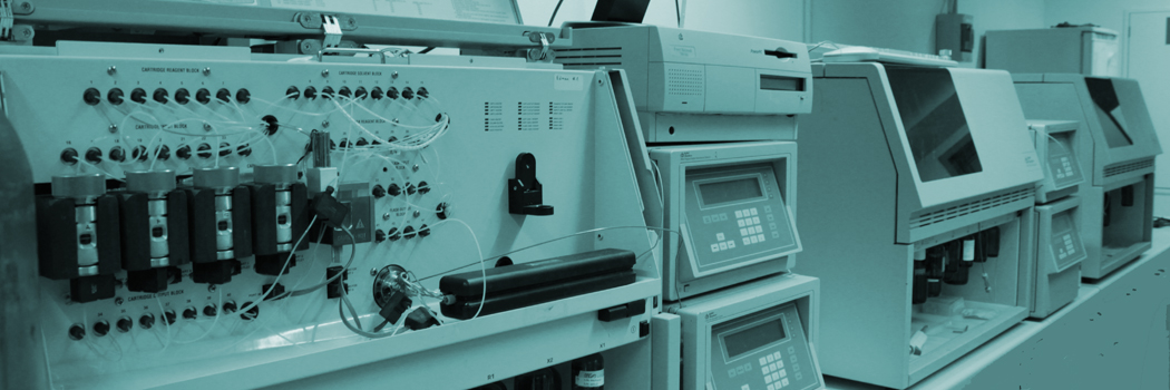
Edman degradation of proteins and peptides is done on ABI's Procise 494 or Procise cLC sequencers. The basic analysis covers the first five amino acids, but up to approximately 40 amino acids can be sequenced. The number of steps (= amino acids) needs to be determined by the client before analysis. The feasibility of longer sequencing runs depends on sample amount, sample quality and the protein's sequence. Our experts can give you guidance on this topic.
Your sample
Sample format: either purified protein (dissolved in or lyophilised from low salt buffer) or an SDS-PAGE band blotted onto PVDF is suitable. At least 25 pmol of protein are required (for five steps), more is highly advisable to improve signal quality. Please note: do no use fast-blotting systems as their proprietary membranes are incompatible with solvents used in Edman chemistry. Please avoid buffers with primary amines for blotting (like Tris and glycine) as they reduce sensitivity and/or give additional signals. An optimised blotting protocol using borate buffer can be found at the bottom of this page.
You receive
a result report of the N-terminal sequence analysis with the detected amino acid sequence and all HPLC chromatograms as a PDF file by email or via one of our SSL and password protected web servers.
How to order
Please contact us first. We can give you some helpful information and send you our protein Edman sequencing sample form. You can also download our N-terminal Edman amino acid sequencing sample submission form here. Then ship the sample together with the completed sample submission form to us.
Sample requirements
| Sample amount | Minimum 25 pmol for five steps, 200 pmol for 15 steps. More sample is highly advisable. Please inquire if you have only smaller sample amounts available. We have a dedicated sequencer for small sample amounts (surcharge applies). |
| Sample type | 1. lyophilized protein/peptide samples 2. liquid protein/peptide sample in 10-100µl volatile solvent like Milli-Q water, isopropanol, acetonitrile. Check whether your sample requires cooled shipping. 3. Protein samples blotted on PVDF membrane (PVDF membrane size: max. 3x6 mm, smaller and more concentrated sample are preferred. The protein band can be stained by Ponceau S, Amido black, Sulforhodamine or Coomassie Brilliant Blue. |
| Sample purity | The sample should be as pure as possible (at least 75-80% purity) and contain only the protein or peptide. Free amino acid, primary amines, SDS, salt, buffer or other contaminants should be as low as possible since the Edman chemistry can be negatively affected. |
| Cysteine modification | Cysteine without special modification can not be detected by N-terminal sequencing. Therefore the sample has to be modified for detection of cysteine. Below you can find a modification protocol. |
| N-terminal blockage | Proteins and peptides which are N-terminally blocked do not have a free N-terminal amino group. Therefore these proteins can not be sequenced. More than 50% of all eukaryont proteins are blocked. On request we can perform deblocking procedures but we need a significant higher amount of protein and the deblocking does not work always, because mostly the type of blockage is unknown. |
| Glycosylation and other modifications | Edman sequencing steps without the detection of an PTH amino acid, reduced peak intensity or altered retention times can be caused by glycosylation, phosphorylation or other modifications. Modified amino acids often cannot be sequenced. We recommend mass spectrometry instead. |
Shipping details
- Please complete our Submission Form for Edman sequencing (pdf, 67 kB) with information about the samples (e. g. sample name, sample type, MW, protein amount, staining method) and the requested number of N-terminal sequencing steps.
- The samples should be in an Eppendorf reaction tube sealed with Parafilm. Protein samples on PVDF membrane can either be shipped as small and dry membrane slides in reaction tubes or as a dried PVDF membrane in plastic foil with enclosed description of the protein bands which should be analyzed. We cut the marked protein band for analysis.
- Liquid samples should be sent in frozen state.
- Lyophilized sample and samples blotted on PVDF membrane can typically shipped at room temperature.
- Send your samples in a padded envelope or in a box and enclose the sample submission form.
- Notify us of your order via Diese E-Mail-Adresse ist vor Spambots geschützt! Zur Anzeige muss JavaScript eingeschaltet sein! or phone, especially if you are shipping international.
Prices (effective 1st February 2025)
| N-terminal protein-/peptide sequencing (includes setup and 5 steps) |
price per sample, standard service (10-15 business days) |
price per sample, |
| 1 sample | 425 EUR | 650 EUR |
| 2 samples | 400 EUR | 600 EUR |
| 3 and more samples | 370 EUR | 550 EUR |
| each further sequencing step (=amino acid) | 50 EUR | 75 EUR |
| setup fee (applies when no sequence information was obtained, e. g. due to N-terminal blocking or insufficient sample amount) |
300 EUR | 450 EUR |
Pricing for analysis of small sample amounts using a Procise cLC sequencer is available upon request.

Our recommendations for protein blotting and staining
Chemical sequencing using Edman's phenylisothiocyanate chemistry targets the primary amino group on the protein's N-terminus. Accordingly buffers containing primary amines like Tris and glycine should be avoided for blotting as they significantly decrease sensitivity.
Semi-dry blotting
- PVDF membrane: Immobilon P membrane or comparable
- Blot buffer: 50 mM sodium borate, pH 9.0 / 20% methanol (HPLC quality) (0.1% SDS can be added to blotting buffer if protein above 40kDa should be sequenced)
- Blotting conditions: 1mA/cm2 PVDF membrane for 2-3 hours at 4°C
- after staining please wash membrane with water (Ponceau S) or 50% methanol/water (Coomassie)
Ponceau S (red) staining
- 0.25 g Ponceau S
- 0.5 mL acetic acid
- ad 50 mL in Milli-Q water
- Procedure:
1. Wash the PVDF blot membrane 2x 3 minutes with plenty Milli-Q water.
2. Stain the PVDF membrane with Ponceau S staining solution for 1-3 minutes.
3. Destain the PVDF blot membrane under visual control with Milli-Q water until protein bands are well visible.
4. Dry the PVDF membrane.
CBB R250 staining
Staining solution (0,1% CBB R250, 10% acetic acid, 40% methanol in Milli-Q water)
-
0.1 g CBB R250
-
40 mL methanol
- 10 mL acetic acid
- ad 10 mL in Milli-Q water
Destaining solution (10% acetic acid, 40% methanol in Milli-Q water):
- 40 mL methanol
- 10 mL acetic acid
- ad 10 mL in Milli-Q water
- Procedure:
1. Stain the PVDF blot membrane for 5 minutes in CBB R250 staining solution
2. Destain the PVDF blot membrane for 3 x 5 minutes with destaining solution under visual control until protein bands are well visible.
3. Dry the PVDF membrane.
Sulforhodamine staining
Staining solution (0,005% sulforhodamine, 0,2% acetic acid, 30% methanol in Milli-Q water)
- 150 mL methanol
- 1 mL acetic acid
- 25 mg sulforhodamine ad 500 mL in Milli-Q water
- Procedure:
1. Wash the PVDF blot membrane 2x 10 minutes with plenty Milli-Q water.
2. Dry the PVDF membrane at room temperature!
3. Stain the PVDF membrane in Sulforhodamine staining solution for 1-2 minutes.
4. Wash the PVDF membrane with Milli-Q water shortly.
5. Dry the PVDF membrane.
Alkylation of Cysteine residues
No signal will be obtained for Cysteine residues when they are not derivatised prior to analysis. Following amino acids will be detected normally, however. Two reagents are recommended for the alkylation of Cysteine thiol groups:
- iodoacetamide, a common reagent which is easy and safe to handle
- 4-vinylpyridine, which gives superior signals in Edman sequencing
See the publication by Sechi et al "Modification of cysteine residues by alkylation. A tool in peptide mapping and protein identification" for more information.


